Biology Notes for Class 12
Chapter 3: Human Reproduction
Chapter Summary
Humans are sexually reproducing and viviparous. The male reproductive system is composed of a pair of testes, the male sex accessory ducts and the accessory glands and external genitalia. Each testis has about 250 compartments called testicular lobules, and each lobule contains one to three highly coiled seminiferous tubules. Each seminiferous tubule is lined inside by spermatogonia and Sertoli cells. The spermatogonia undergo meiotic divisions leading to sperm formation, while Sertoli cells provide nutrition to the dividing germ cells. The Leydig cells outside the seminiferous tubules, synthesise and secrete testicular hormones called androgens. The male external genitalia is called penis.
The female reproductive system consists of a pair of ovaries, a pair of oviducts, a uterus, a vagina, external genitalia, and a pair of mammary glands. The ovaries produce the female gamete (ovum) and some steroid hormones (ovarian hormones). Ovarian follicles in different stages of development are embedded in the stroma. The oviducts, uterus and vagina are female accessory ducts. The uterus has three layers namely perimetrium, myometrium and endometrium. The female external genitalia includes mons pubis, labia majora, labia minora, hymen and clitoris. The mammary glands are one of the female secondary sexual characteristics.
Spermatogenesis results in the formation of sperms that are transported by the male sex accessory ducts. A normal human sperm is composed of a head, neck, a middle piece and tail. The process of formation of mature female gametes is called oogenesis. The reproductive cycle of female primates is called menstrual cycle. Menstrual cycle starts only after attaining sexual maturation (puberty). During ovulation only one ovum is released per menstrual cycle. The cyclical changes in the ovary and the uterus during menstrual cycle are induced by changes in the levels of pituitary and ovarian hormones. After coitus, sperms are transported to the junction of the isthmus and ampulla, where the sperm fertilises the ovum leading to formation of a diploid zygote. The presence of X or Y chromosome in the sperm determines the sex of the embryo. The zygote undergoes repeated mitotic division to form a blastocyst, which is implanted in the uterus resulting in pregnancy. After nine months of pregnancy, the fully developed foetus is ready for delivery. The process of childbirth is called parturition which is induced by a complex neuroendocrine mechanism involving cortisol, estrogens and oxytocin. Mammary glands differentiate during pregnancy and secrete milk after child-birth. The new-born baby is fed milk by the mother (lactation) during the initial few months of growth.
THE MALE REPRODUCTIVE SYSTEM:
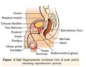

Ø Located in the pelvis region.
Ø Male reproductive system includes
- A pair of testes.
- Accessory ducts.
- Accessory glands.
- External genitalia
Testes:
Ø Located outside the abdominal cavity within a pouch called scrotum.
Ø Scrotum provides low temperature required for spermatogenesis.
Ø Each testis is about 4 to 5 cm length and 2 to 3 cm width.
Ø Each testis has about 250 compartments called testicular lobules.
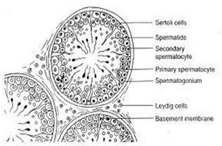
Ø Each lobule contains one to three seminiferous tubules.
Ø Seminiferous tubules lined by male germ cells and Sertoli cells.
Ø Male germ cell undergoes meiosis and produce sperm.
Ø Sertoli cells provide nutrition to the germ cell and the sperm.
Ø In between the seminiferous tubule there is interstitial cell or Leydig cell.
Ø Leydig cells produce testicular hormones called androgen (testosterone).
Accessory ducts:
Ø Includes rete testis, vasa efferentia, epididymis and vas deferens.
Ø Seminiferous tubules open into vasa efferentia through rete testis.
Ø The vasa efferentia leaves the testis and open into epididymis.
Ø The epididymis leads to vas deferens that ascends to the abdomen through inguinal canal and loops over the urinary bladder.
Ø Vas deferens receives a duct from seminal vesicle and opens into the urethra as the ejaculatory duct.
Ø Urethra originates from the urinary bladder and extends through the penis to its external opening called urethral meatus.
Accessory glands:
Ø Includes
- Paired seminal vesicle
- A prostate gland
- Paired bulbourethral gland.
Ø Secretion of these glands constitutes the seminal plasma.
Ø Seminal plasma rich in fructose, calcium, and certain enzyme.
Ø Secretion of bulbo-urethral glands helps in lubrication of penis.
External genitalia:
Ø Penis is the external genitalia.
Ø It is made of special tissue that helps in erection of the penis to facilitate insemination.
Ø The enlarged end of penis is called glans penis.
Ø Glans penis is covered by a loose fold of skin called foreskin.
THE FEMALE REPRODUCTIVE SYSTEM:
Ø Located in the pelvic region of the female.
Ø The female reproductive system includes:
- A pair of ovaries
- A pair of oviduct.
- Uterus
- Cervix
- Vagina
- External genitalia.
- A pair of mammary gland.
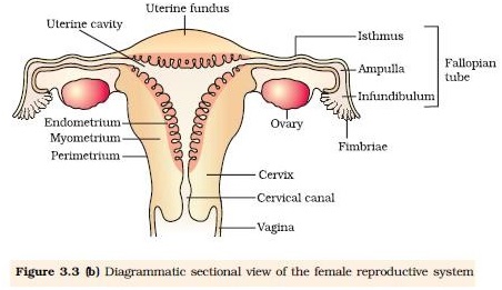
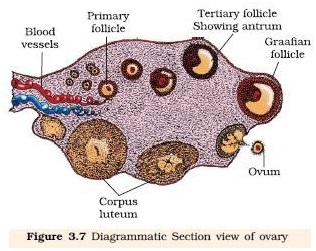
Ovaries:
Ø It is the primary female sex organs that produce the female gamete (ovum).
Ø It also produces several steroid hormones.
Ø The ovaries located in the lower abdomen.
Ø Each ovary is about 2-4 cm in length.
Ø Connected to the pelvic wall and uterus by ligaments.
Ø Each ovary is covered by thin epithelium which encloses the ovarian stroma
Ø The ovarian stroma has two zones
Ø A peripheral cortex.
Ø An inner medulla.
Oviduct:
Ø Oviducts, uterus and vagina constitute the female accessory ducts.
Ø Each fallopian tube is about 10-12 cm long and extends from the periphery of each ovary to the uterus.
Ø Close to the ovary the oviduct has a funnel shaped structure called infundibulum?
Ø The edges of the infundibulum possess finger-like projections called fimbriae, which helps in collection of the ovum after ovulation.
Ø The infundibulum leads to a wider part of the oviduct called ampulla.
Ø The last part of the oviduct is called isthmus which joined to uterus.
Uterus:
Ø It is single and is called womb.
Ø It is inverted pear shaped.
Ø Attached the pelvic wall by ligaments.
Ø The uterus opens into vagina through a narrow cervix.
Ø The lumen of cervix is called cervical canal.
Ø Cervical canal along with vagina form the birth canal.
Ø The wall of the uterus has three layers of tissues
- Perimetrium: external thin membranous.
- Myometrium: middle thick layer of smooth muscles
- Endometrium: inner glandular layer.
Ø Endometrium undergoes cyclical changes during menstrual cycle.
Ø Myometrium exhibits strong contraction during delivery of the baby.
External genitalia:
Ø It includes following structure:
- Mons Pubis: cushion of fatty covered by skin and pubic hair.
- Labia majora: fleshy folds of tissue which extends down from the mons pubis and surrounds the vaginal opening.
- Labia minora: are paired folds of tissue under the labia majora.
- Hymen: the opening of vagina is often covered partially by a membrane called hymen.
- Clitoris: a tiny finger-like structure lies at the upper junction of two labia minora above the urethral opening.
Mammary glands:
Ø Mammary gland consists of glandular tissue and fat.
Ø Glandular tissue of each breast divided into 15-20 mammary lobes.
Ø Mammary lobes contain cluster of cells called alveoli.
Ø The cells of alveoli secrete milk, stored in the lumen of alveoli.
Ø The alveoli open into mammary tubules.
Ø The tubules of each lobe join to form a mammary duct.
Ø Several mammary ducts join to form a wider mammary ampulla.
Ø Mammary ampulla connected to lactiferous duct, through which milk is sucked out.
GAMETOGENESIS: (formation of gametes):
Spermatogenesis:
Ø Formation of sperm from the germ cell in the testes is spermatogenesis.
Ø The process begins at puberty.
Ø Spermatogonia present in the lining of seminiferous tubules undergo mitotic division to increase their number.
Ø Each spermatogonium is diploid (2n) which contain 46 chromosomes.
Ø Innermost layer of spermatogonial becomes larger called primary spermatocyte.
Ø Primary spermatocyte undergoes meiosis-I to form two equal haploid (n) secondary spermatocytes (n).
Ø Each secondary spermatocyte undergoes meiosis-II to form two equal, haploid spermatids.
Ø Each primary spermatocyte produces four spermatids.
Ø Spermatids transformed into spermatozoa (sperms) by the process called spermiogenesis.
Ø The sperm head embedded in the Sertoli cell.
Ø Release of sperm from the seminiferous tubule is called spermiation.

Hormonal control of spermatogenesis:
Ø This process is initiated at puberty due to secretion of gonadotrophins releasing hormone (GnRH)
Ø GnRH secreted form hypothalamus and stimulate anterior pituitary to secrete two gonadotrophins.
- Luteinizing hormone (LH) and
- Follicle stimulating Hormone (FSH)
Ø LH acts on Leydig cells and stimulates synthesis of androgens.
Ø Androgen stimulates spermatogenesis.
Ø FSH acts on Sertoli cells and stimulates spermatogenesis in other ways.
Structure of sperm:
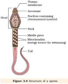
Ø Ultrastructure of sperm consists of a head, neck, a middle piece and a tail.
Ø Whole body of sperm surrounded by plasma membrane.
Ø The sperm head contain an elongated haploid nucleus.
Ø Above the nucleus a cap like structure present called acrosome.
Ø The acrosome contains enzymes which help in fertilization of ovum.
Ø The middle piece contains mitochondria, which provide energy for movement of tail that facilitate sperm motility.
Ø Human male ejaculates 200-300 million sperms during coitus.
Ø 60 percent must have normal shape and size and 40 percent of them must show vigorous motility.
Ø Sperm released from seminiferous tubules enters into accessory ducts.
Ø On their way fluids from seminal vesicle and prostate gland added which collectively called as Semen.
Ø The function of male accessory ducts and glands are maintained by testicular hormone androgen.
Oogenesis:
Ø Formation of a mature female gamete or ovum is called oogenesis.
Ø Oogenesis starts during embryonic stage, 25th week of the fetal age.
Ø Germinal epithelium of ovary divided mitotically to produce millions of gamete mother cell or oogonia.
Ø No oogonia formed or added after birth.
Ø Oogonia enters into meiosis-I and proceeds uptodiakinesis of Prophase-I and get suspended, at this stage called primary Oocytes.
Ø Each primary oocyte surrounded by layers of granulose cells and then called primary follicle.
Ø At puberty only 60,000 to 80,000 primary oocytes are left in each ovary.
Ø After puberty primary follicles get surrounded by more layers of granulosa cells and a new theca to form secondary follicles.
Ø The secondary follicle transformed into tertiary follicle, characterized by a fluid filled cavity called antrum.
Ø The theca layers organized into an inner theca interna and outer theca externa.
Ø During the growth of primary follicle into tertiary follicle during puberty, the primary oocyte restarts its first meiotic division and completes it within tertiary follicle resulting two unequal haploid cells.
- Large haploid cell is called secondary oocyte.
- A tiny cell called first polar body.
Ø The secondary oocyte retains bulk of the nutrient rich cytoplasm of primary oocyte.
Ø The tertiary follicle having secondary oocyte further changes into Graafian follicle.
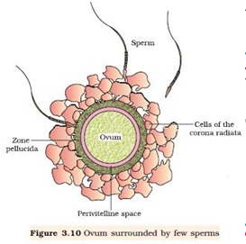
Ø The secondary oocyte surrounded by a new membrane, zonapellucida.
Ø The secondary oocyte undergoes second meiotic division continued upto metaphase-II and get suspended until entry of sperm.
Ø At this stage Graafian follicle releases secondary oocyte from the ovary by the process called ovulation.
Ø On entry of a sperm into the secondary oocytes stimulates it to complete meiosis-II and there is formation of a haploid ovum and a second polar body (n).

GAMETOGENESIS: (formation of gametes):
Ø Reproductive cycle of female primates is called menstrual cycle.
Ø The first menstruation begins at puberty is called Menarche.
Ø Menstrual cycle repeated at an average interval of 28/29 days.
Ø One ovum is released in the middle of each menstrual cycle.
Menstrual cycle has following phases:
i. Menstrual phase:
Ø 1st phase of menstrual cycle.
Ø Menstrual flow occurs.
Ø Lasts for 3-5 days.
Ø Breakdown of endometrial lining and blood vessel.
Ø Mucus and blood comes out through vagina.
Ø It occurs only when ovum released but no fertilization.
Ø Lack of menstruation is the indication of pregnancy.
ii. Follicular phase:
Ø Menstrual phase followed by follicular phase.
Ø Primary follicle becomes Graafian follicle.
Ø Regeneration and proliferation of uterine endometrium.
Ø LH and FSH level increases gradually in follicular phase.
Ø Level of estrogen increases as it is secreted from growing follicle.
Ø It lasts for 5-13 days.
iii. Ovulatory phase:
Ø FSH and LH attain peak level in this period (14th day).
Ø This is called LH surge, which induces rupture of Graafian follicle and release of ovum from the ovary called ovulation.
iv. Luteal phase:
Ø Remaining part of Graafian follicle transformed into corpus luteum.
Ø Coupusluteum produces large amount of progesterone.
Ø Progesterone maintains the uterine endometrium, and prepares it for implantation.
Ø Thickness of uterine endometrium increase in many folds, due to proliferation.
Ø If there is fertilization, corpus luteum grows further and pregnancy continued, menstrual cycle stopped.
Ø In the absence of fertilization corpus luteum degenerates.
Ø Disintegration of endometrium leading to menstruation.
Ø Menstrual cycle ceases around 50 years of age, called menopause.

GFERTILIZATION AND IMPLANTATION:
Ø During copulation (coitus) semen is released by the penis into the vagina is called insemination.
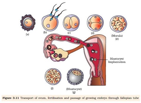
Ø The motile sperm swim rapidly, pass through cervix, uterus and finally reach the junction of isthmus and ampulla (ammpullary-isthmic junction).
Ø The ovum released from the ovary also transported to ampullary isthmic junction where fertilization takes place.
Ø Fertilization only takes place if both sperm and ovum reach ampullary – isthmic junction simultaneously.
Ø The process of fusion of a sperm and ovum is called fertilization.
Ø Acrosome of sperm secretes enzymes helps in penetration into the ovum.
Ø Once a sperm comes contact with the zonapellucida of ovum and induces the changes in the membrane that blocks the entry of additional sperms.
Ø That ensures monospermy and prevents polyspermy.
Ø Only one sperm fertilize with one ovum.
Ø Entry of sperm into the ovum induces the ovum to complete its second meiotic division of secondary oocyte.
Ø Meiosis-II is also unequal cytokinesis resulting production of one large ovum (ootid) and one small second polar body.
Ø Haploid nucleus of sperm fused with the haploid nucleus of ovum to form a diploid zygote.
Sex determination:
Ø Sex of a baby has been decided during fertilization and in the zygote.
Ø Sex is determined by the sex-chromosomes present in gametes.
Ø Human female contain two XX chromosomes.
Ø Human male contain XY chromosomes.
Ø All the female gametes produced with only ‘X’ chromosome.
Ø Sperms produced by male, 50% with ‘X’ and 50 % with ‘Y’ chromosome.
Ø After fertilization zygote either carries XX or XY chromosomes.
Ø Zygote with XX chromosomes develop into female and with XY chromosome develops into male.
Cleavage:
Ø Repeated mitotic division of the zygote without growth resulting a multicellular ball like embryo is called cleavage.
Ø Cleavage starts soon after fertilization.
Ø Daughter cells produced during cleavage are called blastomeres.
Ø The product of cleavage is called Morula, which is 8 to 16 celled.
Ø The morula continues to divide and grow and transformed into blastocyst.
Ø The blastomeres in blastocyst arranged into an outer layer called trophoblast and an inner mass of cells attached to trophoblast called inner cell mass.
Ø Trophoblast cells attached to the endometrium helps development of placenta.
Ø Inner cell mass gets differentiated into the embryo.
Ø After attachment the uterine cells divide rapidly and cover the blastocyst.
Ø Blastocyst completely embedded in the uterine endometrium. This is called implantation.
PREGNANCY AND EMBRYONIC DEVELOPMENT:
Ø After implantation, finger like projections appears on the trophoblast called chorionic villi.
Ø Chorionic villi surrounded by uterine tissue and maternal blood.
Ø Temporary association between the fetal tissue (chorionic villi) and maternal tissue (uterine endometrium) is called placenta.
Function of placenta:
Ø The embryo connected to the placenta by umbilical cord, which transports substances to and from the embryo.
Ø Facilitate transport of oxygen and nutrient from mother to embryo.
Ø Removes CO2 and waste material from the embryo.
Ø Acts as endocrine gland and produces several hormones like:
- Human chorionic gonadotrophins (hCG)
- Human placental lactogen (hPL)
- Estrogen.
- Progesterone
- Relaxin produced from the ovary in the later stage of pregnancy.
Embryonic development:
Ø After implantation the inner cell mass of blastocyst differentiated into an outer layer called ectoderm and an inner layer called endoderm.
Ø Mesoderm differentiated in-between ectoderm and endoderm.
Ø The inner cell mass thus called stem cells, having potency to produce all types of cell, tissues and organs by differentiation.
Organogenesis:
Ø Formation of different organs in the embryo is called organogenesis.
Ø Human pregnancy lasts for 9 months.
Ø After one month of pregnancy heart is formed in the embryo.
Ø By the end of 2nd month the foetus develops limbs and digits.
Ø By the end of 12 weeks (first trimester) most of organ system is formed (limbs and external genitalia are well developed).
Ø First movement of foetus and appearance of hairs observed in 5th month.
Ø By the end of 24th week (2nd trimesters) the body is covered with fine hairs, eye-lids separate, and eyelashes are formed.
Ø By the end of 9 months the foetus is fully developed and is ready for delivery.
PARTURATION AND LACTATION:
Ø The period of pregnancy is called gestation period. (9 months).
Ø Ejection or expulsion or delivery of foetus is called parturition.
Ø Parturition is due to vigorous contraction of uterine Myometrium.
Ø The signal of parturition is originated from the fully developed foetus and the placenta which induces mild contraction of uterus called fetal ejection reflex.
Ø Fetal ejection reflex triggers the release of Oxytocin from pituitary.
Ø Oxytocin induces stronger contraction of uterine endometrium.
Ø Stimulatory reflex continues stronger contraction leads to expulsion.
Ø After delivery the placenta is also expelled out of the uterus.
Lactation:
Ø The mammary gland of the female more differentiated during pregnancy,
Ø Mammary gland starts producing milk towards the end of the pregnancy.
Ø Process of milk production in mammary gland is called lactation.
Ø Milk produced during initial days of lactation is called colostrum.
Ø Colostrum contains several antibodies which provide immunity to the new born baby.
Disclaimer: All contents are originally prepared by Shri K C Meena Ji, Principal, KVS.
Disclaimer: All contents are collected from various sources and updated at this platform to help teachers and students. If content owner (Original Creator) have any objection, Please mail us to info@cbsecontent.com with ID Proof, content will be removed. Thanks and Regards
Notes Station: To Read Click on Title
Book Cart: To Purchase Click on Title


Amazon Affiliate Disclaimer: cbsecontent.com is a part of Amazon Services LLC Associates Program, an affiliate advertising program designed to provide a means for sites to earn advertising fees by advertising and linking to Amazon.in. As an amazon associates we earn from qualifying purchases.
I do not even understand how I ended up here, but I assumed this publish used to be great
I am truly thankful to the owner of this web site who has shared this fantastic piece of writing at at this place.